ULTRASOUND
Ultrasound involves the use of high-frequency sound waves to create images of organs and systems within the body.
How the Test is Performed?
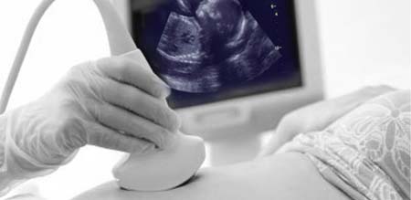
An ultrasound machine creates images that allow various organs in the body to be examined. The machine sends out high-frequency sound waves, which reflect off body structures. A computer receives these reflected waves and uses them to create a picture. Unlike with an x-ray or CT scan, there is no ionizing radiation exposure with this test.
The test is done in the ultrasound or radiology department. You will be lying down for the procedure. A clear, water-based conducting gel is applied to the skin over the area being examined to help with the transmission of the sound waves. A handheld probe called a transducer is moved over the area being examined. You may be asked to change position so that other areas can be examined.
|